Case DVM:
Laura Barnes, DVM, DACVO
Marginal mass affecting the superior nasal eyelid of the OS
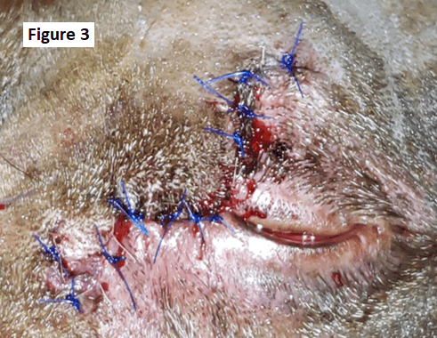
Hershey is a 10.5 year old MC Labrador Retriever that presented to the ophthalmology department for the first time on 11/4/2019 for an enlarging ulcerated cutaneous to eyelid marginal mass affecting the superior nasal eyelid of the OS. He presented to his primary care veterinarian for an initial evaluation of this mass in 10/2019 where 2 days prior his owner noticed that Hershey irritated the mass causing it to bleed. Incisional biopsies were taken at the time of his initial primary care visit. Histopathology revealed an inflamed meibomian gland adenoma carrying a good prognosis with complete surgical removal. Hershey was referred to the ophthalmology department for surgical removal after it doubled in size. Clinical examination revealed allergic conjunctivitis OU, keratitis OS - secondary to mass effect, superior nasal, large ulcerated pink mass OS - 20 mm medial-lateral, 18 mm dorsal-ventral (figure 1.), and nuclear sclerosis OU; Hershey had a normal fundic examination OU. Despite the current histopathology, differentials for the appearance of this mass included squamous cell carcinoma, fibrocarcinoma, mast cell tumor and eyelid hemangiosarcoma/hemangioma.
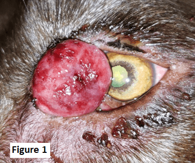
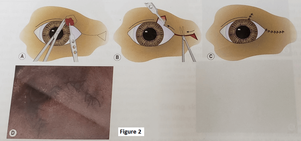
Only approximately 1/3 of the eyelid margin can be removed and closed without the need for eyelid reconstruction. This mass involved a significant amount of superior eyelid margin and given its location we chose a semicircular graft. The semicircular skin grafts are used in dogs and cats for restoration of surgical defects involving 30-60% of the center of upper and lower eyelids. Semicircular grafts utilize both rotational and sliding components (figure 2). Full excision of the superior nasal eyelid mass was performed and a semicircular graft was used to recreate the palpebral fissure and eyelid margin on Hershey (figure 3).
Histopathology revealed severe pyogranulomatous and lymphoplasmacytic inflammation with ulceration of the overlying epidermis that is diffuse with underlying granulation tissue formation along with some necrotic debris. There was no evidence of neoplasia and infectious agents and special staining including PAS, GMS and Fites were negative for infectious agents. This mass likely occurred from previous trauma and/or eyelid infection. It is unknown why there is such a large discrepancy in histopathology results, however lab variation, pathologist training and skills as well as sample size and location may have played a role