

by Laura Barnes, DVM, DACVO
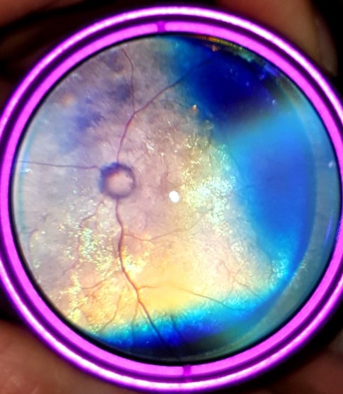
Cessa is a 10 year old FS DSH who presented to the ophthalmology department for acute vision loss. Cessa had an absent menace response OU, normal intraocular pressures and widely mydriatic pupils OU. Her pupillary light reflex was present OU, but sluggish and incomplete. She was losing weight and had a spotty appetite. There was no known travel history. Examination of the anterior segment revealed 1+ aqueous flare. Her fundic examination revealed the following abnormalities.
 |
 |
||
|
Photo A Optic nerve atrophy, tertiary retinal vascular |
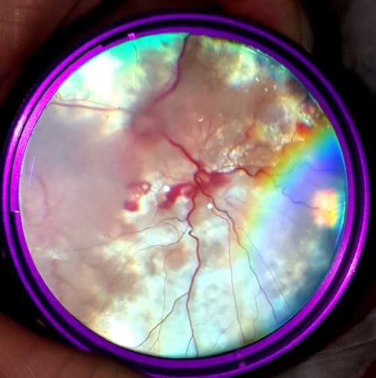
Photo B Optic neuritis, dilated retinal venules with hemorrhage, retinal detachment Subretinal cellular infiltrate |
Complete abdominal ultrasound revealed mild mesenteric lymphadenopathy. Thoracic radiographs revealed a bronchiolar pattern. Based on clinical findings, ultrasound and radiographic findings, we recommended a histoplasmosis titer. Cessa was positive for histoplasmosis. Feline disseminated histoplasmosis is often chronic and insidious with vision loss in some cases being the first presenting signs of a problem. The most common ocular clinical signs include conjunctivitis, chorioretinitis, retinal detachment and optic neuritis. This patient was treated with Sporonox, but never regained vision. She was subsequently lost to follow up.